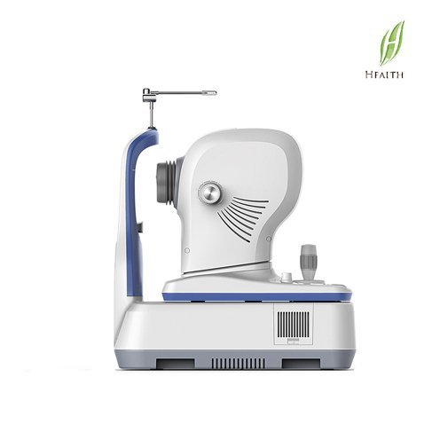
Optical Coherence Tomography (Anterior and Posterior Segment, SLO live fundus image with CE)
FCT-01 is an intelligent SD-OCT and Line Scanning Ophthalmoscope (LSO) combined system with informative outputs, remarkable user interface, exquisite design and reliable quality.
MACULA
Macular HD line
high definition OCT imaging reveals small lesions
Macular Six-line Radial
Having a glimpse of the retina with HD imaging and quick data analysis
Macular Cube
A point-by-point assessment of retinal thickness with a 500*100 dense cube
GLAUCOMA
Glaucoma (Macular)
Glaucoma (Disc)
Informative Report
PERMIUM FUNCTIONS
En-Face Analysis
Network System
Specification:
| OCT Image | |
| Methodology | Spectral domain OCT |
| Optical source | Super luminescent diode(SLD), 840mm |
| Scan speed | 36,000 A-scans per second |
| Axial resolution(optical) | 5 microns( optical), 2.7 microns (digital) |
| Tranverse resolution | 15 microns( optical), 3 microns (digital) |
| A-scan depth | 2.3mm |
| Diopter range | -20 to +20 diopters |
| Scan patterns | Macula:HD line scan(6 or 12mm),3D scan(6*6mm), 6-line scan(6mm) |
| Disc:3D scan (6*6mm) | |
| Anterior segment: 6-line radial scan,HD line scan (6*6mm) | |
| Fundus Imaging | |
| Methodology | Line scanning ophthalmoscope (SLO) |
| Frame rate | 10 fps |
| Minimum pupil diameter | 3.0mm |
| Field of view | 50 degrees |
| Software analysis | |
| Macula | Retina thickness analysis; Progressive analysis |
| 3D view; En-face analysis | |
| Glaucoma | Ganglion cell analysis; Cup-disk analysis |
| RNFL analysis; Ou comparative analysis | |
| progression analysis | |
| Anterior segment | Manual Measurement; Corneal thickness analysis |
| Others | DICOM conformance; Remote viewer software avilable |
| Electrical and physical | |
| Weight | 29kg |
| Dimension | 450(L)*250(W)*45(H)mm |
| Source voltage | AC 100 0 240V |
| Frequency | 50 Hz – 60 Hz |
| Power input | 90VA |


