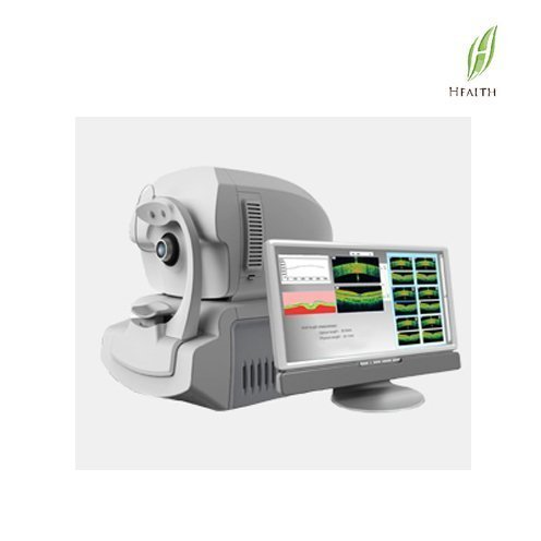
FCT-03 not only have day after anterior segment OCT and OCT data acquisition and analysis function, more will depend on optical measurement function integrated into an organic whole. Can complete the cornea, the anterior segment structure such as corner fault morphology observation, in section structure after retina and other qualitative and quantitative observation, and the axial length, anterior chamber depth, corneal curvature, and to measure the data feature set in one, improve the amount of information obtained single collection, comprehensively improve the efficiency and the accuracy of clinical diagnosis.
Character:
1. Simultaneous imaging of both the anterior and posterior segments
2. It uses the latest patented technology to realize simultaneous imaging both the anterior and posterior segment.
3. During the inspection, FCT-03 do not need to add or change anterior segment lens, just scan once time, you can get clear images both anterior and posterior.
4. It could make the patient more comfortable and need the least time, so as the doctor’s working efficiency is also improved.
5. Accurate ocular axial length measurement
6. Compare with the traditional ultrasonic ocular axial length measurement, the coherent light biological measurement technology which FCT-03 adopts could ensure the precision and accuracy of the measurement. When doctor get OCT images,the ocular axial length value is also gainable at the same time.
7. Posterior segment functions
8. Macular lutea and optic disk scan and analysis
9. 3D ILM,IS/OS,RPE topographic map
10. RNFL scan analysis, glaucoma follow-up analysis and two eyes comparison functions
11. Anterior segment function
12. Cornea, chamberangle, iris, crystalline lens scan Cornea curvature and thickness topographic map
Specification:
| Signal Type | Internaland external fixation |
| Light Source | Photo scattered from tissues |
| Optical power | Super Iuminescent LED,840nm |
| Anterior and posterior axial resolution | <=0.75mw |
| Posterior lateral resolution | 5um |
| Anterior lateral resolution | 10um |
| Cormeal thickness measurement precision | 15um |
| Corneal curvature measurement precision | 20um |
| Ocular axial length measuring range | 0.1mm |
| Ocular axial length measurement precision | 14-40mm |
| Anterior chamber depth measuring range | 0.02mm |
| Imaging function | 1.5-6.5mm |
| Optical measurement | Simultaneous imaging both the anterior and posterior segment |
| Scan modes | Ocular axial length, anterior chamber depth, cornealcurvature, corneal diameter(white to white),crystal surface curvature and etc |
| Scan rate | Macular iutea, opticdisk, one line and 12 lines in cornea, one line in anterior chamber angle, ocular axial length, anterior chamber depth and crystalline lens thickness |
| Acquisition time | 36,000A- scan/SEC |
| Scan Depth | 70 pictures per second |
| Oculi funds diopter adjustment range | 2mm in tissue |
| Fixation modes | -20D-+20D |


