Product Abstract:
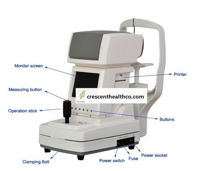
Sciencetera’s innovative DSP engine for FRE-12 Ref-keratometer controls the entire measurement process including capture of measurement signal as well as, the data process and the display.So FRE-12 enables to provide stable measurement data and fine pupil images for you.
Features:
■ When operate FRE-12, you can expect a high quality and best customer service.
■ Auto-Starting
■ As soon as FRE-12 is properly aligned to each eye, this function initiates the measurement process and generates the measurement results automatically.
■ Automatic Fogging
■ The FRE-12 incorporates an infinity scene diopters of fog to control accommodation.
■ Instuinctive, Easy of use UI.
■ Its users can enjoy convenient and comfortable operation due to easy of use UI.
■ HR LCD ( High Resolution Color LCD Monitor )
■ 0.3 Mega Pixel 6.4 inches TFT Color LCD provides fine and large iris image with color alignment marks.
■ Automatic PD Measurement
■ FRE-12 automatically calculates pupilary distance for both near and far vision.
Other measurements
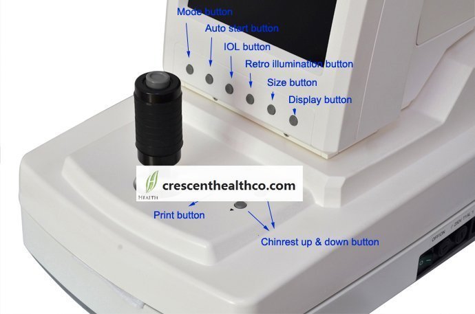
IOL, PD, Pupil diameter, CLBC, Ret-illum, etc.
Alignment marks
Move closer to the patient
Move further to the patient.
Incorrect collimation
Specifications:
| Refractometer mode | ||||
| Sphere | -25.00D~+22.00D | |||
| Cylinder | 0~+/-10D | |||
| Axis | 1~1800 | |||
| Keratometer mode | ||||
| Corneal curvature | R5.0mm~R10mm | |||
| Corneal refraction | 67.5D~33.7D | |||
| Corneal astigmatism | 0~+/-10D | |||
| Axis angle | 1~1800 | |||
| Other measurement mode | ||||
| Pupil distance | 0~85mm | |||
| Pupil diameter | 0~12mm | |||
| CLBL | R5.0mm~R10mm | |||
| IOL | OK | |||
| Ret-illum | OK | |||
| Others | ||||
| Measuring start | Auto /Manual | |||
| Display | 6.4 inches 0.3M TFT color LCD | |||
| Alignment | Color display | |||
| Printer | Built in printer | |||
| Output | RS232C /USB( option port ) | |||
| Power supply | AC110~240Vac 50/60Hz 100VA | |||
| Dimension | 242( w ) x467( d ) x465( h ) | |||
| Weight | 16.5kg | |||
Retro-illumination
Retro-illumination is another form of indirect viewing, where the light is reflected off the deeper structures,
such as the iris or retina. This method is useful for examining the size and density of opacities, but not their location.


